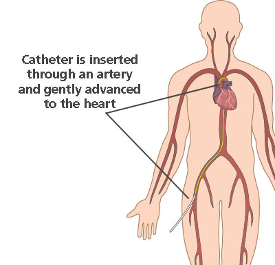
Catheter Advanced From the Right Femoral Vein
An arterial catheter for invasive hemodynamic monitoring was inserted into the right femoral artery and an Arrow-Howes Large-Bore Multi-Lumen Central Venous Catheter 12 Fr. Both ports had good venous blood return.

Biosensors Free Full Text Ai Enabled Ultrasound Guided Handheld Robotic Device For Femoral Vascular Access Html
The long sheath was advanced this length over the guide wire.
. Through the right femoral vein a catheter was advanced to superior vena cava. Place the probe in a sterile sheath with coupling gel inside. The right femoral vein was dilated.
Despite looping of the catheter ablation and termination of atrial flutter were performed successfully. The femoral vein is accessed using the Seldinger technique with a 19- or 21-gauge needle. Sterile gel can be applied to the outside of the protective covering.
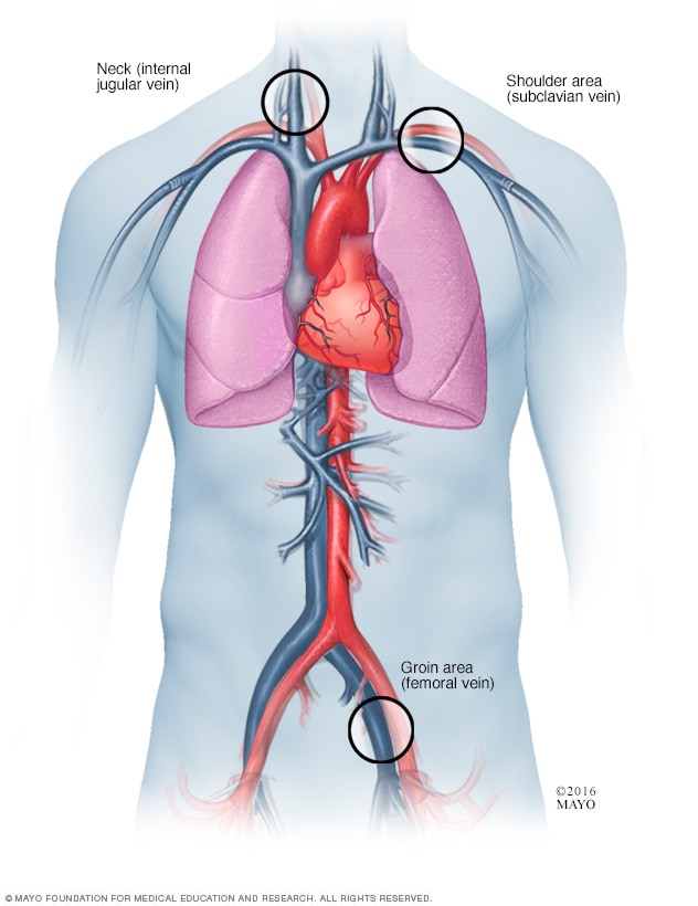
A central venous catheter CVC is an indwelling device that is peripherally inserted into a large central vein most commonly the internal jugular subclavian or femoral and advanced until the terminal lumen resides within the inferior vena cava superior vena cava or. The guidewire was advanced smoothly. Placing the patient in a reverse Tren delenberg position engorges the femoral vein and may aid in visualization.
Both ports were flushed with saline and heparin. Using a volcano intravascular ultrasound catheter IVUS is performed from the femoral vein to the heart. Question 1 1 out of 1 points Catheter advanced from the right femoral vein into the left and right pulmonary artery.
These catheters are advanced via the femoral veins in the groin to the right andor left atrium. The hook needle was taken out. In front of you.
Attempted to advance guidewire from right femoral vein to superior vena cava. In the operating room the venous catheter was replaced by tubing from the port system kit. Both ports had good venous blood return.
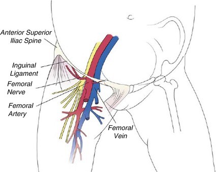
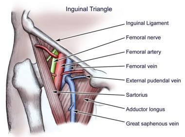
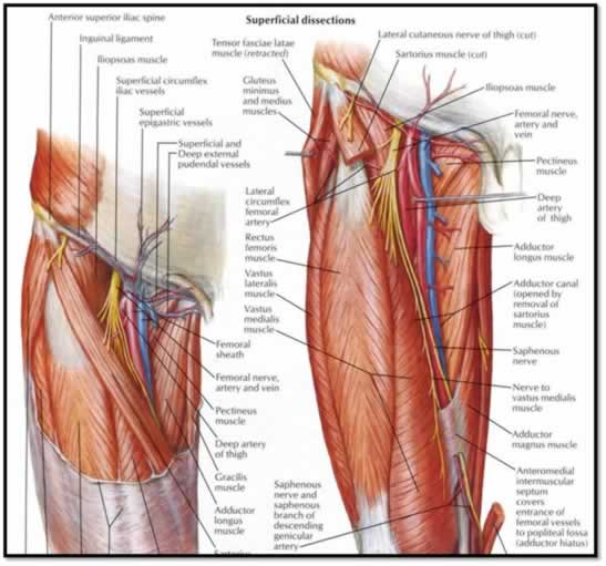
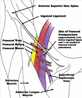
The catheter was further negotiated into the right lung lower lobe. Contrast is injected and imaging performed 75833-59. Pearls A helpful mnemonic to remember the location of the femoral vein is VAN V ein A rtery N erve which indicates the order in which the three structures are encountered from.
A catheter was inserted via femoral access without complications. When simultaneous adrenal venous sampling is to be performed bilateral femoral punctures or two unilateral femoral vein punctures are made. Cobara catheter were subsequently advanced into the right atrium through the femoral vein.
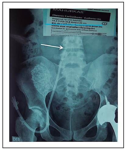
Ablation for Ventricular Tachycardia Ventricular tachycardia usually presents in. An 115 French 16-cm temporary dialysis catheter with a straight extension was advanced to the iliac vein through the right femoral vein over the guidewire using the Seldinger technique without difficulty. An 115 French 16-cm temporary dialysis catheter with a straight extension was advanced to the iliac vein through the right femoral vein over the guidewire using the Seldinger technique without difficulty.
The Kumpe catheter is then advanced beyond the heart and into the right subclavian vein. The catheter was then gently. From a right femoral vein approach a catheter is advanced into the right ovarian 36011-59 left renal and the left ovarian veins 36012.
Once the vein is cannulated the catheter is threaded over the needle into an intraluminal position and the needle is withdrawn leaving the catheter in place. The catheter was further negotiated into the right lung lower lobe. We decided to use right common femoral venous entry and discussed the situation with the patient.
Encountered great resistance at junction of right femoral vein and inferior vena cava. Significant findings are recorded. The guidewire tract was then dilated and then a triple lumen venous catheter was inserted over the guidewire and advanced into the right femoral vein.
These images show renal vein reflux into massively dilated venous structures supplying numerous pelvic varicosities on the left side. Multiple attempts at locating the CS os with contrast injection through a Swartz Guiding sheath SL2 St Jude Medical were also unsuccessful. An experienced physician inserted a right femoral catheter Dual lumen catheter 10 French in diameter and 15 cm in length with some difficulty and resistance on advancement of the guide wire and the catheter itself.
The catheter was secured to the skin. Guidewire advanced into superior vena cava. This is the first report of an inferior-to-superior approach for ablation of atrial flutter in the absence of the perihepatic IVC.
Before insertion of these sheaths the distance from the femoral puncture to the mid sternum was measured to estimate the distance to the right atrium. Orient the probe so that the patients right is on the right side of the screen. The large bore needle was then withdrawn.
Pulmonary angiography performed in all locations including radiologic supervision and interpretation. Catheter advanced from the right femoral vein into the left and right pulmonary artery. Subsequently the guide wire was retracted behind the fix curve of the Cobara catheter and the catheter bent spontaneously in the absence of supporting guide wire following retraction.
To enable right-sided CVC placement in patients with TCVO an inside-out access IOA approach was established at 3 vascular access centers in Europe involving use of a novel IOA device advanced from the right femoral vein. Adrenal vein catheterization for venography and sampling is done from a right femoral vein approach. Agilis deflectable St Jude were then advanced through both femoral veins over a guide wire to the right atrium.
The patient provided additional informed consent to proceed. An ablation catheter could be advanced through the right femoral vein reaching the right heart via vena azygos and SVC. He underwent an invasive cardiac electrophysiology EP study.
Dilator placed prior also could not navigate this point. Pulmonary angiography performed in all locations including radiologic supervision and interpretation. The catheter was further negotiated into the right lung lower lobe.
An 0035-inch 0088-cm Amplatz super-stiff wire is then placed via the catheter. A guidewire was passed through the large bore needle into the right femoral vein and advanced. Pulmonary angiography performed in all locations including radiologic supervision and interpretation.
20 cm catheter length Arrow International Inc Reading PA. Catheter advanced from the right femoral vein into the left and right pulmonary artery. Via the right femoral vein the authors were unable to cannulate the coronary sinus CS with a deflectable decapolar catheter.
CVCs cause thoracic central vein occlusions TCVOs more often than right-sided internal jugular catheters.
2
Cardiac Catheterization Heart Care Intermountain Healthcare
The Usual Vascular Access Springerlink

References In Better With Ultrasound Chest
Arterial Line Placement Background Indications Contraindications

Distal Superficial Femoral Vein Cannulation For Peripherally Inserted Central Catheter Placement In Infants With Cardiac Disease Richter 2016 Congenital Heart Disease Wiley Online Library

1 Catheters Are Inserted Within The Heart Using The Right Femoral Vein Download Scientific Diagram
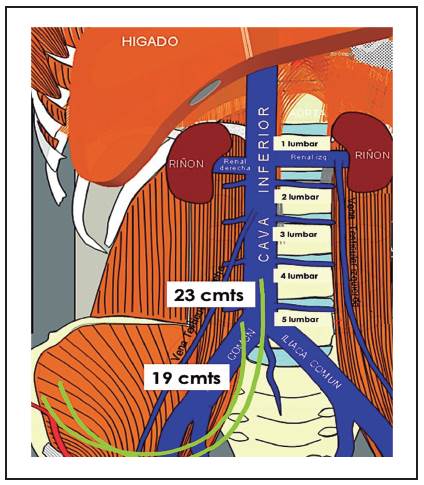
Tuneled Catheters In Femoral Vein Does The Length Makes Any Difference

Surface Modification Strategies For Hemodialysis Catheters To Prevent Catheter Related Infections A Review Balikci 2021 Journal Of Biomedical Materials Research Part B Applied Biomaterials Wiley Online Library
Procguide Femoral Central Line
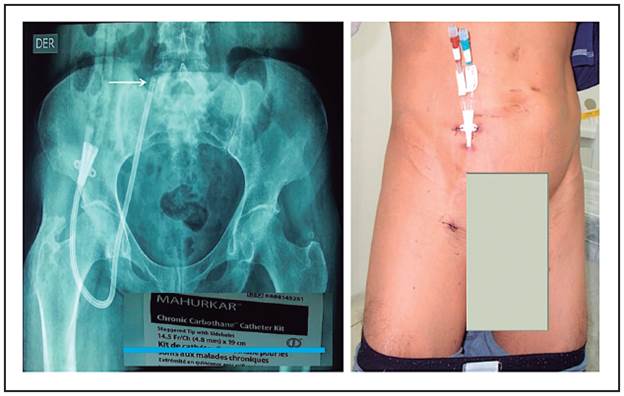
Tuneled Catheters In Femoral Vein Does The Length Makes Any Difference

Femoral Central Lines Vascular Wellness

9 Femoral Venous Catheterization Download Scientific Diagram
![]()
A Catheter Blue Arrow Insertion Into The Femoral Vein Red Arrow Download Scientific Diagram
Pulmonary Vein Isolation Mayo Clinic
Femoral Central Venous Access Technique Femoral Vein Cannulation Complications
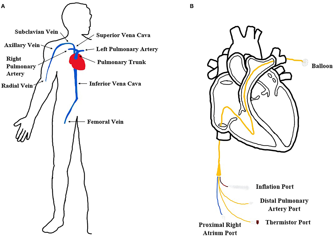
Frontiers An Inexpensive Cardiovascular Flow Simulator For Cardiac Catheterization Procedure Using A Pulmonary Artery Catheter Medical Technology
Tuneled Catheters In Femoral Vein Does The Length Makes Any Difference

Tandemheart A Multi Staged Access Cannula Is Inserted Via The Femoral Download Scientific Diagram


Comments
Post a Comment